[ad_1]

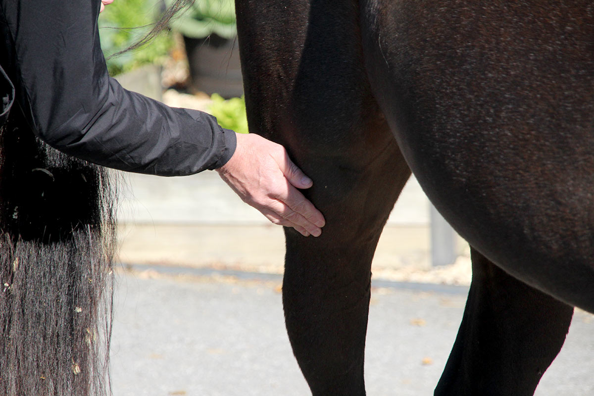
From hip to hoof, rather a lot can go fallacious in a horse’s hind limb. A mess of small bones, ligaments, tendons, and muscle groups, work in tandem to propel a horse ahead, soar, flip shortly, and even simply enable them to stay standing whereas they relaxation. The stifle is a fancy joint within the horse’s higher hind-limb and performs an vital position in his capability to maneuver with clean impulsion and stand for lengthy intervals of time.
“Stifle lameness is comparatively widespread, occurring in all sorts of horses and disciplines,” says Ellen Regulation, DVM, ECVDI, a big animal resident within the Diagnostic Imaging Clinic on the College Animal Hospital, in Uppsala, Sweden. “It’s particularly widespread in leaping, barrel racing, slicing, and high-level dressage horses.”
Stifle Anatomy
Whereas usually regarded as a single joint, the stifle consists of three joints: two femorotibial joints and the femoropatellar joint. Throughout the stifle, the tip of the femur (thigh bone) divides into two fistlike, spherical bulbs of bone. The entrance a part of these “fists” consists of the medial (internal) and lateral (outer) trochleas, which don’t immediately bear any weight and don’t articulate (transfer) with the tibia (the internal of the 2 bones that extends down from the knee to the hock). Somewhat, the trochleas articulate with the patella (a small, cartilage-covered bone colloquially known as the kneecap), forming the femoropatellar joint. The medial and lateral condyles make up the underside of the fists, which articulate with the highest floor of the tibia, bearing all of the horse’s weight.
A flat disc of versatile, sturdy fibrocartilaginous tissue lies between every condyle and the tibia, offering cushioning throughout weight bearing. This joint is subsequently known as the femorotibial joint. To complicate issues barely, the medial and lateral femorotibial joints don’t talk (join in order that fluid can movement between the joints) in lots of horses, making the stifle three separate joints.
Every joint within the stifle connects to vital soft-tissue constructions, such because the collateral ligaments, muscle groups, tendons, and meniscal fibrocartilages and their related ligaments that connect the menisci (the cartilaginous discs between the femur and the tibia that facilitate frictionless stifle joint motion) to each the tibia and femur.
Sue Dyson, MA, VetMB, PhD, an impartial guide from the U.Ok., describes how these constructions work in movement. “Within the femoropatellar joint, the patella glides up and down over the cranial (towards the top) side of the trochleas,” she says. “The three patellar ligaments come up from the bottom of the patella and are successfully the insertions of the quadriceps muscle, an vital extensor muscle of the stifle.”
The three patellar ligaments course medially, laterally, or straight down and converge on the entrance of the tibia.
Frequent Stifle Pathology
Dyson says widespread circumstances inflicting lameness within the stifle of horses embrace:
- Osteoarthritis (OA) of the femorotibial joint, with the medial compartment affected extra generally than the lateral;
- Meniscal damage both with or with out simultaneous damage of the cranial or caudal (towards the tail) meniscal ligaments;
- Upward fixation/delayed launch of the patella, occurring extra usually in younger fairly than mature horses; and
- Persistent osteochondrosis of the femoropatellar joint, extra usually within the lateral fairly than medial trochlea. Ostochondrosis is a comparatively widespread developmental abnormality characterised by a defect in cartilage and the underlying bone. Osteochondrosis. Affected joints usually have effusion and may end up in lameness.
“The medial femorotibial joint and associated constructions, such because the medial collateral ligament and medial meniscus, are mostly injured,” says Regulation. “That is because of the biomechanics and loading of the limb throughout train.”
A Nearer Have a look at Stifle Lameness in Horses
Osteoarthritis of the femorotibial joint
The femorotibial joints bear weight immediately, so trauma, irritation, and different elements that result in OA trigger lameness by step by step breaking down the cartilage on these weight-bearing surfaces.
“OA might be secondary to meniscal damage, anatomical variants/developmental anomalies, flattening of the medial femoral condyle, concavities within the articular floor of the distal (additional away from the physique) side of femur, and subchondral bone cysts,” says Dyson.
Remedy for any such OA resembles others and may embrace oral nonsteroidal anti-inflammatory medicine (NSAIDS, e.g., phenylbutazone) or joint injections with corticosteroids, orthobiologics comparable to interleukin-1 receptor antagonist protein/IRAP, or artificial hydrogels.
Meniscal damage both with or with out simultaneous damage of the cranial or caudal meniscal ligaments
“Meniscal accidents and accidents to their related ligaments are widespread,” says Dyson. “The meniscal cartilages transfer somewhat throughout flexion and extension, however these actions are restricted by the meniscal ligaments. They’re subsequently topic to massive masses, shear forces, and torsional pressure that predisposes them to damage.”
Horses can maintain differing types and places of meniscal tears, and just some might be addressed arthroscopically.
Upward fixation/delayed launch of the patella
“Delayed launch might be extra widespread in all sorts of horses,” says Dyson. “On this case the affected limb(s) is (are) not caught in extension, however the motion of the patella is jerky, not clean.”
Veterinarians and house owners may solely observe delayed launch when the horse strikes laterally (facet to facet, e.g., asking the horse to step away from you on the bottom) or when transitioning from trot to stroll. It happens repetitively however usually intermittently. “This situation unquestionably causes persistent ache and lowered efficiency,” Dyson provides.
Upward fixation is far simpler to acknowledge as a result of the patella does lock—close to the tops of the trochleas—with the limb in extension. This situation principally self-rectifies or might be by pushing the horse backward.
Conservative care entails bettering the horse’s muscling by instituting a managed train program and making certain the horse’s acceptable vitamin for his age and workload. When this situation causes persistent lameness and femoropatellar joint effusion (swelling), veterinarians may decide to transect (reduce) the medial patellar ligament, additionally referred to as ligament splitting.
“If the medial patellar ligament is cut up in a number of websites, it’s nonetheless intact, so the keep equipment is totally intact,” says Dyson, referring to a community of muscle groups, tendons, and ligaments that permits a horse to face with minimal effort. “Transection is finished much less generally now due to potential postoperative issues and is adopted by restore of the ligament in a lengthened, thickened type.”
Persistent osteochondrosis of the femoropatellar joint
On this painful situation the patella applies strain to the femoral condyles, inflicting painful cartilage dysfunction and subchondral bone harm. Dyson says unfastened items of cartilage can grow to be indifferent and progressively ossify, leading to movable items within the joint. On account of its poor high quality, the cartilage may fail to correctly take up shock for the underlying bone.
Remedy often consists of surgical procedure, though very early situations of the situation in younger foals may self-repair. “Persistent instances in mature sports activities horses are tough to handle,” Dyson says.
Deal with the Patellar Ligaments
Regulation lately studied the patellar ligaments in additional element, hoping to grasp if horses expertise persistent ache originating from these three ligaments and the infrapatellar fats pad (a comfortable tissue pad behind the kneecap)—a situation much like “jumper’s knee” in human athletes.
After scanning the 116 horses’ patellar ligaments on ultrasound, Regulation and colleagues discovered a big proportion of horses in full work had ultrasonographic adjustments within the patellar ligaments.
“These adjustments have been similar to lesions described in earlier literature, comparable to bleeding and irritation within the ligaments (desmitis),” says Regulation. “This highlights the significance of thorough scientific examination and blocking when investigating stifle lameness to verify that findings on ultrasound are clinically important.”
Take-Dwelling Message
Stifle lameness is widespread in horses and is usually a results of extra than simply these 4 circumstances; nevertheless, they’re probably the most incessantly recognized. Veterinarians ought to carry out a cautious scientific examination, block nerves with native anesthetics, and conduct imaging utilizing radiography, ultrasonography, or computed tomography (CT) to establish the underlying reason for the lameness.
[ad_2]
Supply hyperlink
